AMH as a Marker of Ovarian Reserve in Perimenopause
AMH predicts ovarian reserve more reliably than FSH during perimenopause.

Perimenopause is not an event but a stage, and a long one at that: for most women it runs four to seven years, though for some the hormonal transition stretches out to a decade. The most useful single marker for tracking where a woman stands inside that stretch is anti-Müllerian hormone, or AMH, because it declines in a steady, roughly predictable way as the ovarian follicle pool shrinks. That makes it a clearer signal than the two hormones most people associate with perimenopause testing, FSH and estradiol, both of which swing unpredictably from one cycle to the next. This piece walks through what AMH actually measures, why its biology sets it apart from reactive hormones, and what a declining AMH trajectory means in practical terms for a woman trying to understand her own transition.
The reproductive research community uses a framework called STRAW+10 to stage this transition with some precision: early transition (stage −2), late transition (stage −1), the final menstrual period (stage 0), and early postmenopause (+1a). A single biological fact underlies all of these stages: ovarian reserve depletes continuously from birth, producing the changes each stage tracks. Ovarian reserve, the remaining pool of viable follicles, is finite from birth and depletes continuously across a woman's life. The rate and timing of that depletion vary considerably between individuals, which is why measuring reserve, rather than guessing at it from symptoms or age alone, matters. Many women reach their mid-40s experiencing mood changes, disrupted sleep, and cycle shifts with no real number to tell them where they are in the process, because the right test hasn't been ordered or interpreted with any context.
What follicular depletion does to the hormonal environment
Follicles do not disappear in a smooth, linear fashion. As they become fewer and less responsive to signaling, the hormonal outputs that depend on them shift in an erratic, sometimes jarring pattern rather than a gradual descent.
Estrogen, produced by the ovaries, reaches receptors throughout the brain, the cardiovascular system, bone, skeletal muscle, and metabolic tissue. When its output starts fluctuating unpredictably instead of following its usual cyclical rhythm, every one of those systems feels it, and the effects appear as mood changes or sleep disruption long before anyone thinks to check a hormone panel. Progesterone tells a somewhat different story: because its synthesis depends on ovulation actually occurring, it tends to fall earlier and more consistently than estrogen does. That mismatch creates a period of relative estrogen dominance in the early perimenopausal years, and progesterone's decline carries its own consequences, since it plays a direct role in bone density, cardiovascular protection, and cognitive function.
FSH behaves differently still. As follicles grow more resistant to signaling, the brain compensates by producing more FSH to try to recruit them into development. FSH rises, but the rise is reactive, a response to follicular resistance rather than a steady readout of anything. AMH sits in a different category. It is produced by growing follicles and plays a regulatory role, making certain follicles more resistant to FSH so that only the most viable ones get selected for ovulation each cycle. As the total follicle pool shrinks, AMH output shrinks along with it, in a direct, proportional way. Estrogen and FSH react to what is happening cycle by cycle. AMH reflects how many follicles are actually left. That distinction runs through the rest of this discussion.
The symptom picture that often arrives before any test is ordered
A study published in Menopause, the official journal of The Menopause Society, co-authored by Mayo Clinic and Flo Health, surveyed 17,494 women across 158 countries and asked what symptoms they associated with perimenopause. Hot flashes topped the list at 71%, followed by sleep problems at 68% and weight gain at 65%.
But that is not what women in perimenopause report experiencing most often. Fatigue, exhaustion, irritability, low mood, sleep disruption, and anxiety dominate the actual lived experience, with mood and energy complaints often overshadowing the vasomotor symptoms everyone expects. That gap between expectation and reality carries real clinical weight. A woman who shows up describing anxiety, brain fog, or fragmented sleep often gets evaluated for thyroid issues, depression, or stress before anyone considers ovarian aging as a possibility.
Part of the problem is timing. STRAW+10 lists vasomotor symptoms, the hot flashes and night sweats, as characteristic of late transition, stage −1. By the time those symptoms appear in force, the transition is already substantially underway. The earlier signals, the fatigue and mood shifts, arrive first and get missed most often. No single symptom profile applies universally. Severity, timing, and which symptoms dominate vary meaningfully across women, shaped in part by ethnic, cultural, and nutritional factors. Symptoms alone, in other words, are not a reliable map of where someone stands. That unreliability is why hormonal markers matter, and why the marker chosen matters too.
Why FSH is an unreliable guide to where a woman stands in the transition
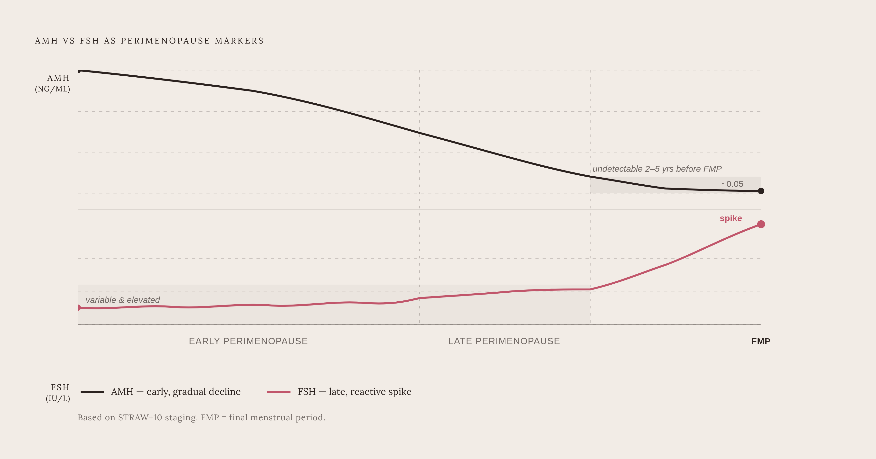
FSH remains the most commonly ordered marker in this context, and there's a reasonable case for it: persistently elevated FSH above 25 to 30 IU/L does correlate with menopausal-range hormonal changes. The trouble is what "persistently" is doing in that sentence.
FSH is a reactive signal. The brain produces more of it in response to follicles failing to respond, which means it rises sharply only near the end of the transition rather than tracking it throughout. Worse, FSH levels can swing considerably week to week, even day to day, during perimenopause, and a result can drop back into premenopausal range on a given draw for no clear reason. This variability is well documented enough that it has become a standard clinical caveat: don't trust a single FSH number.
STRAW+10 reflects this directly. For stage −1, elevated FSH above 25 IU/L is listed only as a supportive criterion, not a primary one. For stage −2, the earlier stage, FSH is described merely as "variable and elevated," without even that specific threshold attached. The primary criteria driving staging are menstrual pattern changes, not FSH values. Consider a woman of 47 with irregular cycles, hot flashes, and broken sleep whose FSH comes back within normal range. That result does not clear her of anything. She may simply have been tested between surges, and the test's silence does not cancel the clinical picture in front of her. Estradiol carries a similar limitation, fluctuating dramatically across the cycle and depending heavily on which cycle day the blood was drawn, which makes a single measurement difficult to interpret with any confidence during a transition defined by irregular cycles. Both markers share the same structural flaw: this flaw causes them to capture what the reproductive axis happens to be doing at the moment of the draw, rather than how much reserve actually remains, as shown by their dependence on cycle timing rather than on reserve itself.
What AMH measures and why its biology sets it apart as a marker
AMH comes from granulosa cells inside growing follicles. That single fact separates it from FSH and estradiol in a fundamental way: AMH is a direct output of follicle activity, not a downstream response to follicles failing.
Its regulatory job, reducing early follicles' sensitivity to FSH so only the strongest ones proceed to ovulation while the rest undergo atresia, means AMH governs which follicles get selected, rather than simply reflecting the outcome after the fact. Because it's produced continuously by every growing small follicle, not just the one dominant follicle chosen each cycle, its level tracks the size of the whole follicular pool. That is ovarian reserve in its truest functional sense, not a proxy for it.
The behavioral difference that matters most in practice is that AMH does not cycle meaningfully across the menstrual month. It's essentially cycle-day independent, which means it can be drawn on any day without the tight Day 3 to Day 5 timing window that FSH and estradiol testing require. For a woman whose cycles have already gone irregular, which is most women by the time they're asking these questions, that timing requirement is often impossible to meet anyway. AMH sidesteps the problem. Inhibin B appears in STRAW+10 as another supportive criterion alongside AMH, though it has a less established research base as a longitudinal predictor. AMH has the deeper research base as a longitudinal predictor, and that steady, non-reactive decline over years, rather than a reactive spike near the end, is what makes it genuinely useful for mapping where someone sits in the transition. The next section gets into what that decline actually looks like in numbers.
How AMH declines across the transition
Estimated mean AMH concentration at the time of menopause runs around 0.05 ng/mL, compared with 1.36 ng/mL in women still having regular menstrual cycles. Estimated mean AMH concentration at the time of menopause runs around 0.05 ng/mL, compared with 1.36 ng/mL in women still having regular menstrual cycles. That is not a subtle shift. It unfolds over years rather than days, and longitudinal studies show AMH typically becomes undetectable somewhere between two and five years before the final menstrual period, signaling the approaching end of the transition well before cycles actually stop.
A 2025 review in PubMed examining AMH's role in ovarian insufficiency and menopause prediction found that AMH declines steadily with age and functions as a reliable predictor of menopausal timing. One study cited in that review, looking at women aged 35 to 39 who were still premenopausal, found a median time to menopause of 9.94 years for those with AMH below 0.20 ng/mL, versus 13.01 years for those above 1.5 ng/mL. That's a spread of roughly three years driven entirely by baseline AMH, which is meaningful information for anyone doing family planning or long-range health counseling.
None of this should be read as precision it doesn't have. Relying on AMH alone to predict an exact age of menopause carries real uncertainty, with confidence intervals ranging from two to twelve years for women under 40. AMH points a direction; it does not mark a date on a calendar. Still, it outperforms FSH for long-range prediction specifically because it declines gradually across years while FSH only rises sharply near the very end of the process, giving AMH a longer runway as a usable signal. Clinically, combining AMH with other assessments tends to produce a more complete picture than relying on FSH or estradiol alone.
The limits of AMH and their effect on interpretation
None of this makes AMH a diagnostic switch. The 2025 PubMed review is explicit that AMH's reliability for pinpointing menopause is inconsistent, particularly in younger populations or when trying to nail down an exact onset date. It's a predictor, and a good one, but not a verdict.
The confidence intervals bear repeating here because they matter for how the number gets used: two to twelve years for women under 40 is too wide a range to build precise family-planning decisions on without pulling in other clinical context. And the picture gets complicated in the other direction for women with polycystic ovary syndrome. Elevated AMH in PCOS is associated with a delayed onset of menopause, roughly two years longer on average (mean age 51.4 in PCOS cases versus 49.7 in healthy controls, a 2025 review found), which means a high AMH reading needs to be read against a woman's reproductive history, not treated as a universal reassurance.
External factors complicate things further. Pelvic radiotherapy studies found that AMH dropped earlier than FSH in treated patients, with 39.1% of patients meeting AMH-based criteria for premature ovarian insufficiency compared with 34.8% by FSH criteria. That gap shows AMH can register ovarian damage from treatment, not just natural aging, which is useful information but also means a low AMH reading always needs context about what might have caused it. There is also no single "low AMH" threshold that applies across every age. A level that looks reassuringly normal at 42 might indicate accelerated depletion if it showed up at 35. AMH works best as one piece of a larger panel rather than a number read in isolation, which is precisely the position STRAW+10 gives it: a supportive criterion alongside inhibin B. That's not a weakness particular to AMH so much as a reflection of how the field as a whole thinks about ovarian aging: no single marker tells the whole story.
Why serial AMH testing reveals what a single result cannot
A single AMH draw tells you roughly where someone stands. It cannot tell you which direction they're moving or how fast, and that second piece of information affects how the results should be acted on.
The rate of AMH decline varies significantly across age groups, and each woman's trajectory is shaped by her own individual biology. Population averages describe the general curve, but they cannot place any individual woman on it with confidence from a single point. That is the entire argument for testing more than once. Longitudinal AMH measurement allows for a more individualized prediction of menopause timing, which in turn supports better counseling on both family planning and the management of the health issues that tend to cluster around the menopausal transition, bone density and cardiovascular risk chief among them.
Consider two women with the same current AMH value on paper. One has held roughly steady across two annual tests; the other has dropped substantially over that same year. Their numbers might look identical sitting side by side on a population chart, but they are in genuinely different clinical positions, and only the serial data reveals that difference. AMH answers a question that FSH, estradiol, and progesterone cannot, which is where reserve is headed rather than what the reproductive axis is doing this week. Together, these markers answer more than any single one does alone. But raw numbers, even trended over time, do not interpret themselves. Age, symptom history, cycle patterns, and the rest of the hormone panel all have to enter the reading for the data to become something actionable rather than just a number on a lab report. Understanding what a given trajectory means in practice is really a question of what to do with it, which is where the conversation with a clinician comes in.
How to use AMH results to have a more productive conversation with a clinician
AMH does not diagnose perimenopause on its own. Mayo Clinic is clear on this point: no single test or symptom confirms that the transition has started. Clinicians weigh age, menstrual history, and reported symptoms or body changes together, and AMH is one input into that broader assessment.
What AMH contributes is a read on where ovarian reserve stands relative to age-expected norms, and, with repeat testing, a sense of direction that symptoms and cycle tracking alone simply cannot supply. For a woman experiencing mood changes, disrupted sleep, and cycle irregularity while her FSH comes back "normal," AMH is often the test that catches what FSH, reactive and late-arriving by nature, misses. It's worth asking specifically for AMH as part of a panel that also includes FSH, estradiol, and progesterone, since each of these markers is answering a slightly different question, and the combination builds a picture no single result can offer on its own.
STRAW+10 itself carries known limitations for certain groups, since women on hormonal contraceptives, women with PCOS, and women who've had a hysterectomy or endometrial ablation all require a clinician who understands how those factors distort the standard staging picture. That expertise matters as much as the lab result itself. It also helps to ask that results get documented in a way that allows future tests to be compared directly against a personal baseline, since that is what turns a handful of scattered snapshots into a trajectory a clinician can actually act on.
Perimenopause remains a years-long transition that gets under-explained and under-diagnosed far more often than it should. A woman who arrives at an appointment holding AMH data and some sense of her own trajectory is in a fundamentally stronger position to advocate for herself than one relying on symptoms alone to make her case.
Sources
- The role of anti-Müllerian hormone: insights into ovarian reserve, primary ovarian insufficiency, and menopause prediction - PubMed
- Role of anti-Müllerian hormone in predicting premature ovarian insufficiency after pelvic radiotherapy - PubMed
- The role of anti-Müllerian hormone: insights into ovarian reserve, primary ovarian insufficiency, and menopause prediction | Endocrine | Springer Nature Link
- journals.lww.com
- Frontiers | Anti-Müllerian hormone beyond an ovarian reserve marker: the relationship with the physiology and pathology in the life-long follicle development
- academic.oup.com
- asrm.org