Bone Density Loss Timeline During the Perimenopausal Transition
Bone density drops sharply in a three-year window around menopause, not gradually with age.
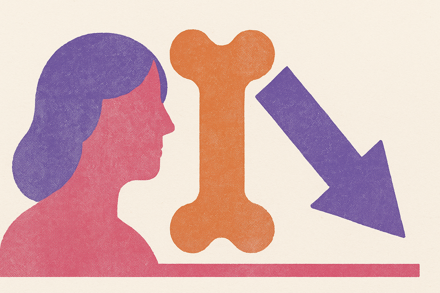
Bone density loss during perimenopause does not creep. It concentrates into a short, measurable window around the final menstrual period, and understanding exactly when that window opens, how fast it moves, and what drives it gives women a far more useful picture of their own risk than the vague warning that bone "thins with age."
Perimenopause and the distinct bone-loss window
The common assumption treats bone loss as background noise, a slow erosion that tracks quietly alongside wrinkles and graying hair, picking up speed in some vague way as the decades pass. That assumption does not hold for the menopausal transition. The data describe something closer to a cliff than a slope: a period of stability, followed by a compressed phase of rapid density loss, followed by a slower plateau. Before perimenopause begins, bone density in most women holds steady. It is not already declining at a lower rate and simply accelerating. One might argue that if bone loss were truly gradual, the timing of intervention would hardly matter. But if the loss is concentrated into a defined window, timing becomes the whole strategy. A woman who believes bone loss is a slow drip has no particular reason to act in her late 40s. A woman who understands that a sharp, time-limited phase of loss is coming has a reason to prepare for it before it starts. That distinction, between a gradual slide and a bounded window, is the premise the rest of this piece works from.
Rapid bone loss and the STRAW+10 staging system
Clinicians map the menopausal transition using a framework called STRAW+10, which divides the years around menopause into discrete stages: the early menopausal transition (stage −2), the late menopausal transition (stage −1), and the first year following the final menstrual period (stage +1a). The bone-loss window this piece is concerned with sits across the boundary between the late stages, roughly where stage −1 gives way to stage +1a. Stage −2 itself is defined clinically by a concrete marker: a difference of seven days or more between consecutive menstrual cycle lengths. An irregular cycle, in other words, functions as the first clinical signal of the transition, long before most women would think to connect it to their skeleton. STRAW+10 has a known limitation: symptom onset frequently arrives before a woman's cycles meet the formal criteria for perimenopause. A woman can already be in a state of real hormonal flux while her periods still look regular enough to escape the staging definition. That matters for bone specifically: density loss may begin before any staging criteria would classify a woman as perimenopausal.
When rapid bone loss begins, how fast it runs, and when it slows, according to SWAN data
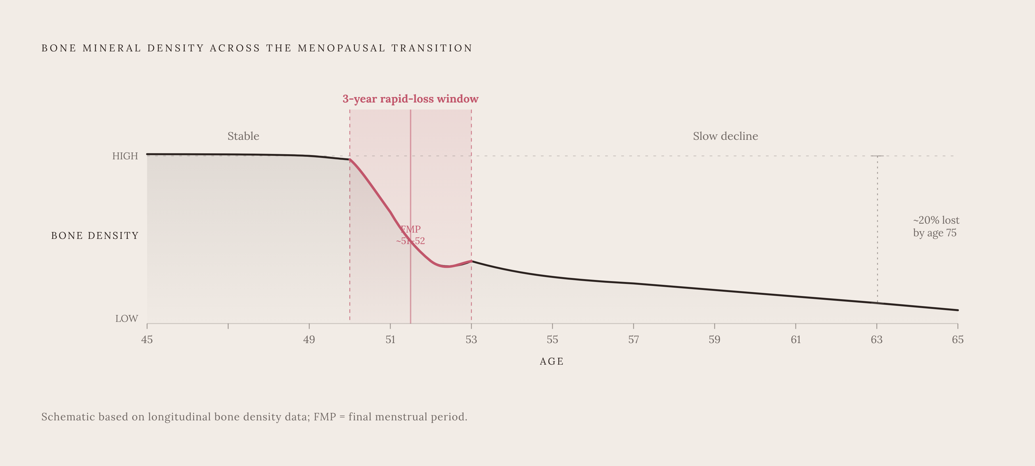
A large longitudinal cohort study followed women prospectively from before their cycles changed through the postmenopausal years, and it offers the clearest longitudinal map available of when rapid bone loss starts and when it stops. The rapid phase begins approximately one year before the final menstrual period. It runs for roughly three years. During those three years, women lose bone density at a substantial rate each year, with the spine losing faster than the hip, and the cumulative effect over a decade amounts to a meaningful deficit in skeletal mass. Before this phase opens, density holds steady. After the three years pass, the rate of loss slows again. The window does not represent a new, permanent rate of decline but a finite, high-velocity phase that eventually gives way to a calmer trajectory. A 2026 StatPearls clinical review places this phase in a longer arc, finding that between menopause and age 75, a woman may lose roughly a fifth of her total body bone mineral. The rapid three-year phase documented by SWAN is the frontloaded portion of that longer decline, the part where the damage accumulates fastest and where the stakes of inattention are highest. SWAN's data carry another finding that reframes the whole conversation around risk: faster loss during the transition predicted more fractures later, independent of how much bone a woman started with. Pace, not just starting point, carries prognostic weight. A woman who starts with strong bones but loses them quickly during this window is not protected by her starting density the way intuition might suggest.
Not all women lose at the same rate: who faces the steepest trajectory
The three-year average SWAN describes is exactly that, an average, and averages conceal a meaningful split in experience. The PeKnO Study identified a subgroup researchers labeled "fast bone losers," defined as women who lost a substantial proportion of bone mineral density within two years. Roughly a quarter of participants in that study met the threshold. A quarter is a large enough share to treat as a routine clinical concern. What pushes a woman into that faster group? Several risk factors compound each other rather than acting independently: early menopause before age 45 (which extends the total time spent in a low-estrogen state), low body weight, cigarette use, a sedentary lifestyle, long-term corticosteroid use, and a family history of fractures. A review focused on precision bone imaging notes that women who reach menopause before 45 face a longer cumulative stretch of estrogen deficiency, and that extended exposure shifts their entire bone-loss trajectory earlier and steeper than the average case. The three-year window SWAN describes functions as a population-level picture, not a personal guarantee. A woman carrying several of these risk factors at once may be losing bone faster, earlier, and with no outward sign of it. The hormonal mechanism behind the loss deserves closer attention.
Why estrogen loss alone does not fully explain bone loss timing
Estrogen has long been the explanation offered for perimenopausal bone loss, and the explanation is accurate as far as it goes. Estradiol, the dominant form of estrogen during the reproductive years, suppresses the activity of osteoclasts, the cells responsible for breaking down old bone tissue. When estradiol falls, and falls erratically rather than in a smooth line, bone breakdown begins to outpace new bone formation, and density drops. That mechanism is incomplete, and newer research points to at least two additional, independent drivers: follicle-stimulating hormone (FSH) and the loss of ovulatory progesterone. Why would a hormone like FSH, typically discussed only in the context of fertility, have anything to do with bone? Ramírez Stieben and colleagues, publishing in Calcified Tissue International, found that FSH was independently associated with bone mineral density at the lumbar spine, while estradiol was the stronger predictor at the femoral neck. The two hormones appear to act on different skeletal sites through different pathways, which suggests FSH is not simply a passive marker that rises as the ovaries age. The same research identified a threshold near 15 mIU/mL, above which the relationship between FSH and bone shifts, implying a non-linear effect where skeletal impact changes at a specific point. The PeKnO Study contributes the second piece of this more complex mechanism: progesterone. As ovulatory cycles declined across the study period, bone density loss accelerated in direct correlation with that decline. Progesterone is produced only during ovulatory cycles, and its apparent protective role against bone loss disappears as cycles become anovulatory through perimenopause, independent of what estrogen is doing. Whether FSH directly activates osteoclasts on its own, or whether it mainly signals the broader process of ovarian aging already underway, is not a settled question. The clinical community has not resolved that debate, and the honest position is to treat FSH as a second meaningful signal and not yet a fully understood cause.
What hormone testing can and cannot tell you about bone risk
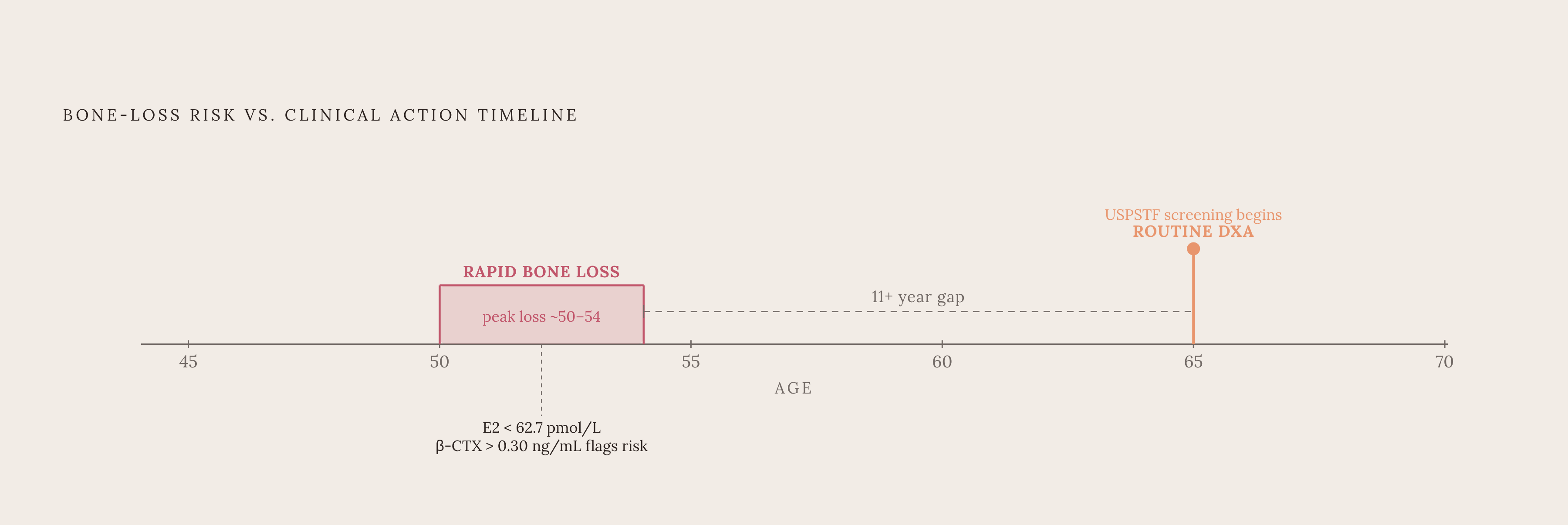
Given three hormones with independent and partially distinct effects, what can a blood panel actually tell a woman about her bone risk during this transition? The relevant markers are estradiol (E2), FSH, and bone turnover markers, and the three together paint a more useful picture than any single value read in isolation. The European Society of Endocrinology's Clinical Practice Guideline, issued in October 2025, states that biochemical testing is not necessary for diagnosing or managing perimenopause in women older than 45. That guidance answers a narrower question than the one this piece is asking: it addresses whether hormone testing is needed to confirm that a woman is in the transition, not whether hormone testing is useful for stratifying her bone risk once she is in it. Those are different clinical questions, and the guideline should not be read as discouraging the kind of targeted testing described here. Feng and colleagues, publishing in Frontiers in Endocrinology, found significant correlations between E2, a bone resorption marker called β-CTX, and T-scores, and found that combining E2 with β-CTX produced a markedly more sensitive and specific prediction of bone loss than either marker measured alone. That study produced two concrete reference points, an E2 threshold of 62.7 pmol/L and a β-CTX threshold of 0.30 ng/mL, giving clinicians and patients fixed numbers to flag high-risk status. FSH adds a third dimension to this picture. SWAN's data found FSH more informative than E2 for identifying which women were more or less likely to begin losing bone, particularly at the lumbar spine, though further research is needed before FSH stands on its own as a clinical bone biomarker. A single blood draw, though, only ever captures one moment. Tracking E2, FSH, and turnover markers across multiple draws over time reveals a trajectory, and a woman whose FSH is climbing while her E2 falls across sequential tests occupies a meaningfully different risk position than one whose values are holding steady, even if any one draw from each woman looked similar on its own.
Why DXA catches bone loss late in perimenopausal women
Dual-energy X-ray absorptiometry, or DXA, remains the gold-standard imaging tool for diagnosing osteoporosis, and nothing here disputes that role once a diagnosis needs confirming. Timing is the problem. DXA registers bone loss only after a substantial portion of trabecular bone has already deteriorated, which makes it poorly suited to catching the rapid perimenopausal window while it is still in progress. Feng and colleagues state this directly in their 2025 paper: DXA is not sensitive enough to detect early bone loss, and an easier, more sensitive method is urgently needed to identify high-risk women before the loss becomes irreversible. That statement functions as the stated motivation behind their work developing biochemical thresholds, not a passing caveat. Current screening guidance compounds the gap. The USPSTF's January 2025 recommendation calls for DXA screening in women 65 and older, and for postmenopausal women younger than 65 only if they carry one or more risk factors for osteoporosis. A perimenopausal woman in her late 40s, already inside or approaching the rapid-loss window, is not automatically flagged for a scan under that framework. SWAN's three-year rapid-loss window runs through approximately age 50 to 54 for many women, centered on a final menstrual period that occurs at a mean age of roughly 51 to 52. That is more than a decade before routine DXA screening begins at 65. The standard screening pathway, in other words, is structurally out of step with the period when the most bone is being lost. Biochemical markers, E2 combined with β-CTX, alongside FSH tracked over time, offer a way to generate an earlier signal during the window itself, well before a DXA scan would show anything a clinician could act on.
How perimenopausal bone loss gets missed in clinical practice
The DXA timing gap does not exist in isolation. It sits inside a broader pattern in which perimenopause itself receives little structured attention in routine clinical care, and many women pass through the bone-loss window with no information about it. A national survey by Biote, titled "Perimenopause Focus," found that fewer than half of clinicians initiated conversations about perimenopause with their patients, and only a small fraction of women felt adequately informed when their symptoms first began. Bone health, several steps removed from the symptoms that typically prompt a visit in the first place, rarely enters a conversation that a clinician does not start. The same survey documented a related misdiagnosis pattern: over a third of women reported feeling misdiagnosed when they sought care during this period, with mood, anxiety, and depression frequently treated as the presenting problem instead. If the transition itself often goes unnamed, its downstream effects on the skeleton stand even less chance of coming up. A woman noticing irregular cycles and asking what is happening to her body is, by these numbers, statistically more likely to walk away with a psychiatric diagnosis than a conversation about where her bone density is heading. That is not a failure of any one clinician so much as a gap in how the transition gets trained and recognized across the system. Which raises a fair question for any woman now reading this with her own cycle length, her own family history, and her own set of risk factors in mind: if the window will not be flagged for her automatically, what does she have to lose by asking the questions herself, before the three years she cannot get back have already passed?
Sources
- Perimenopause & Bone Density: The 3-Year Window
- A threshold of β-CTX (0.3 ng/mL) with low estradiol identifies high-risk perimenopausal women for bone loss: a cross-sectional study
- How Menopause Affects Bone Density: What You Should Know
- Perimenopausal Bone Loss Is Associated with Ovulatory Activity—Results of the PeKnO Study (Perimenopausal Bone Density and Ovulation)
- A threshold of β-CTX (0.3 ng/mL) with low estradiol identifies high-risk perimenopausal women for bone loss: a cross-sectional study
- Perimenopausal Bone Loss Is Associated with Ovulatory Activity—Results of the PeKnO Study (Perimenopausal Bone Density and Ovulation)
- Bone Mineral Density Changes during the Menopause Transition in a Multiethnic Cohort of Women - PMC
- Executive summary: stages of reproductive aging workshop (STRAW) - Fertility and Sterility