Which Hormones Should Be Tested Together in a Perimenopause Panel
Testing multiple hormones together reveals what a single FSH test cannot.
Perimenopause remains one of the more chronically under-recognized transitions in women's health, with symptoms frequently waved off as stress, anxiety, or "just getting older." Part of the reason is structural: the standard approach to testing relies on one hormone, checked once, interpreted with nothing around it for context. Most women in their 40s who raise concerns with a doctor get a single FSH test, and that one number is asked to carry the entire diagnostic weight. But FSH does not hold still long enough for one draw to mean much. A snapshot tells you almost nothing about the trend it is supposedly part of, and treating it otherwise is where a lot of missed diagnoses start.
This is not a fringe objection. NICE guidelines state that FSH testing is not even required to diagnose perimenopause in women over 45 who present with typical symptoms, and yet the single FSH test remains the default in most clinical settings. That gap between guideline and practice is the whole problem in miniature. Estradiol fares no better tested alone. Without FSH, LH, and a progesterone reading timed to the right point in the cycle, an estradiol number says nothing about what is happening with androgens, adrenal hormones, or a thyroid overlap that can produce the identical symptom picture. One might argue any hormone test beats none. True as far as it goes, but a number that cannot be read in context is closer to noise than signal, and that noise is what gets women's symptoms waved off a second time.
How perimenopause changes the hormonal landscape over time
Perimenopause is the years-long hormonal transition that precedes menopause, typically starting sometime in a woman's 40s, though it can begin as early as the mid-30s in some cases. Median onset is around age 47, and the average age of menopause in the United States is 52 https://www.testing.com/tests/female-hormone-panel/. Premature menopause, onset before age 40, affects roughly 1% of people, a reminder that "typical" describes a median, not a guarantee anyone can bank on https://www.testing.com/tests/female-hormone-panel/.
The clinical framework for staging this transition is called STRAW+10, updated in 2011 by an international consortium and still treated as the standard for describing where a woman sits in reproductive aging. It draws on menstrual cycle characteristics alongside FSH, AMH, inhibin B, estradiol, and antral follicle count to place a woman at a specific stage. In the early transition, Stage -2, cycle length shifts persistently by more than 7 days, FSH starts climbing, and progesterone is already on its way down. By the late transition, Stage -1, cycles stretch to 60 days or more for two cycles running, and the swings grow sharper and less predictable. Once a woman reaches postmenopause, FSH and LH sit chronically elevated while estradiol and progesterone drop below the reference ranges seen in the reproductive years.
Here is what actually makes a single-hormone test unreliable: the shape of the whole arc, not just the noise around any one point on it. Progesterone is usually the first hormone to fall, often years before estrogen shows any real decline, and that early drop drives a good deal of the early symptom burden women report. Estrogen itself does not decline in a straight line. Estrogen runs high and erratic for a stretch before gradually falling, so a woman can spend a chunk of perimenopause fighting symptoms of estrogen excess before the low-estrogen symptoms she was told to expect ever appear. Meanwhile FSH and LH climb as the pituitary gland works harder to push a slowing ovary into producing hormone, essentially shouting louder because the ovary is listening less. Androgens, testosterone and DHEA among them, decline on their own slower timeline, gradual rather than a sharp drop. That slide runs partly independent of what estrogen and progesterone are doing. A panel has to track it on its own terms instead of assuming it moves in lockstep with everything else.
The essential core: FSH, estradiol, progesterone, and LH tested together
FSH comes from the pituitary and stimulates the ovaries, rising as ovarian reserve declines. A value above 25 IU/L in the early follicular phase, or in a woman with amenorrhea or infrequent periods, points strongly toward perimenopause, though pinning down one universal cutoff is harder than it sounds because the number moves so much https://academic.oup.com/ejendo/article/193/4/G49/8281862 https://lolahealth.com/blogs/longevity/perimenopause-blood-test-guide. In full menopause, FSH sits consistently above 30 IU/L https://lolahealth.com/blogs/longevity/perimenopause-blood-test-guide. A 2025 guideline is blunt: FSH cannot be read on its own, and estradiol has to be measured alongside it just to make the FSH value mean anything. Because FSH swings so widely, a second measurement 4 to 6 weeks later is often warranted, especially where pregnancy is still a live consideration. A peer-reviewed review from September 2026 in the Journal of Clinical Medicine argues FSH may be doing more than regulating the ovaries at all. FSH receptors have turned up in bone, fat tissue, the brain, blood vessels, immune cells, and the kidneys, which suggests the hormone might work as a broader signal of aging across the body. That is an open question, not settled science, but it reframes why FSH deserves more than a single passing test, especially one that can read normal one day and menopausal-range a fortnight later.
Estradiol, the dominant and most biologically active form of estrogen, is what FSH cannot supply on its own. Low estradiol drives the vasomotor symptoms most people associate with menopause: hot flashes, night sweats, disrupted sleep. Irregular estradiol rhythms can appear years before a final period, an early tell that ovarian function is already shifting. But estradiol without FSH and LH sitting next to it is only half a picture. Together estradiol, FSH, and LH show what the ovaries are actually producing and how hard the brain is working to compensate for the shortfall.
Progesterone is the hormone most often mistested, full stop, and it is usually the first to decline, sometimes years ahead of estrogen. Low progesterone causes a long list of early perimenopausal complaints: disrupted sleep, anxiety, irritability, heavier periods, shortened cycles. Timing decides everything here. It needs to be drawn at the mid-luteal phase, roughly 7 days after ovulation, around day 21 in a standard 28-day cycle, though that day shifts if the cycle itself runs longer or shorter. Test on the wrong day and the number turns meaningless. A day-21 reading below 16 nmol/L suggests ovulation is not happening consistently, and a luteal-phase reading below 10 ng/mL points to anovulation or a corpus luteum not doing its job https://www.testing.com/tests/female-hormone-panel/ https://lolahealth.com/blogs/longevity/perimenopause-blood-test-guide. The luteal-phase reference range itself climbs to 10 to 25 ng/mL after ovulation https://holistic.health/journal/hormone-panel-explained-tests. This is where misdiagnosis happens most: draw the test on the wrong day, or draw it just once, and a genuinely low progesterone level reads as "normal," while the symptoms it would have explained get chalked up to something else.
LH, also from the pituitary, triggers ovulation and rises as estrogen output falls with age. In perimenopause and beyond, LH secretion turns irregular and elevated, with occasional surges, and chronic elevation becomes the norm once menopause arrives. Read individually, FSH, estradiol, progesterone, and LH each tell a partial story. Read as a set, they start to describe an actual trajectory. In the early follicular phase, levels below 100 pmol/L suggest low ovarian output, with a premenopausal reference range of 25–75 pg/mL.
Why thyroid markers belong in every perimenopause panel
The overlap between thyroid dysfunction and perimenopause runs wide enough to cause real diagnostic confusion. Fatigue, weight gain, anxiety, brain fog, thinning hair, irregular cycles, heart palpitations, low mood: all of it appears in both conditions, sometimes in nearly identical combinations. Up to 1 in 4 women investigated for menopause symptoms turn out to have a thyroid disorder, either instead of perimenopause or layered on top of it, with real consequences from the overlap https://lolahealth.com/blogs/longevity/perimenopause-blood-test-guide. That is not a coincidence, and treating it as one is how women end up medicated for the wrong problem.
TSH alone will not catch this. It misses subclinical hypothyroidism, and it misses early autoimmune thyroiditis before the number has drifted out of range. Free T4 is the main circulating thyroid hormone, and a low FT4 paired with an elevated TSH confirms hypothyroidism. Free T3 is the active form, and testing it catches the women whose T4-to-T3 conversion is faulty, something TSH alone will never reveal. TPO antibodies identify autoimmune thyroiditis, and they can turn positive well before TSH itself drifts out of range, which makes them an early warning rather than a confirmatory afterthought. A woman can have a TSH sitting comfortably in range while her Free T3 conversion is genuinely poor, and the symptoms that result just get filed under perimenopause. TSH is the screening marker, with a normal range of 0.27–4.2 mIU/L, though many women feel best below 2.5.
Androgens, AMH, and SHBG: what the expanded panel reveals that the core cannot
AMH, or Anti-Müllerian Hormone, is the most stable marker of ovarian reserve available. On its own it does not diagnose perimenopause. But low AMH paired with low estradiol and high FSH builds a picture considerably more convincing than any one marker standing alone.
Testosterone, both total and free, and SHBG round out the androgen side of the panel. This decline runs its own steady, independent slope alongside the estrogen drop, not caused by it. SHBG governs how much testosterone is actually free and biologically available. It shapes what the total testosterone number even means. Elevated SHBG, driven by oral estrogen use or by hyperthyroidism, drags down free testosterone and free estradiol at the same time. Calculating the Free Androgen Index catches functionally low testosterone even in women whose total testosterone looks unremarkable on paper, exactly the case a total-only test would miss. Symptoms tied to androgen decline, reduced libido, fatigue, muscle loss, a flattened mood, overlap heavily with low-estrogen symptoms, and they get missed constantly when estradiol is the only hormone anyone thinks to check.
DHEA-S adds the adrenal layer. It comes almost entirely from the adrenal glands and works as a precursor hormone for both estradiol and testosterone. A low DHEA-S reading can point toward adrenal insufficiency, though it should never be read apart from cortisol. As ovarian hormone production winds down through perimenopause, the adrenal glands take on a larger relative share of the work. This marker earns a spot on the expanded panel instead of staying optional. Put the full set together, estradiol, progesterone, testosterone, FSH, LH, DHEA-S, and AMH, and each contributes a distinct piece: reproductive health, metabolic function, overlapping conditions like PCOS sitting alongside perimenopause itself. Total testosterone and SHBG together allow the Free Androgen Index calculation, FAI = 100 × total testosterone ÷ SHBG, which is how functionally low testosterone gets caught even when the total number looks fine. Median testosterone declined from 0.56 nmol/L (ages 40–44) to 0.42 nmol/L (ages 55–59), while androstenedione declined by 51% and DHEA by 33% from age 40–44 to 65–69, an independent, gradual trajectory alongside the estrogen decline.
Metabolic markers: why cardiovascular, blood sugar, and bone-related tests complete the panel
Estrogen does more than regulate the reproductive cycle. It protects blood vessels and shapes lipid metabolism, and as estrogen output declines, LDL cholesterol and triglycerides tend to climb in response. A baseline lipid panel drawn during perimenopause gives a woman and her doctor something concrete to track that shift against, and it opens the door to catching a bad trend early rather than after it has already done damage.
Blood sugar regulation follows the same logic. Estrogen offers some protection against insulin resistance, and the hormonal shifts of perimenopause carry a documented link to rising metabolic syndrome risk. An HbA1c test screens for diabetes risk and, just as important, sets a baseline before that metabolic risk has room to accelerate. Vitamin D belongs on the list too, not as an afterthought tacked onto the end of a requisition form. Levels tend to fall around and after menopause, partly because estrogen helps activate vitamin D in the body, and low vitamin D speeds up bone density loss, one of the more serious long-term consequences of declining estrogen. A CBC and a CMP screen for blood cell abnormalities, electrolyte balance, and kidney and liver health, and abnormal liver or lipid values can change which treatment options are even on the table.
Timing testing across the menstrual cycle
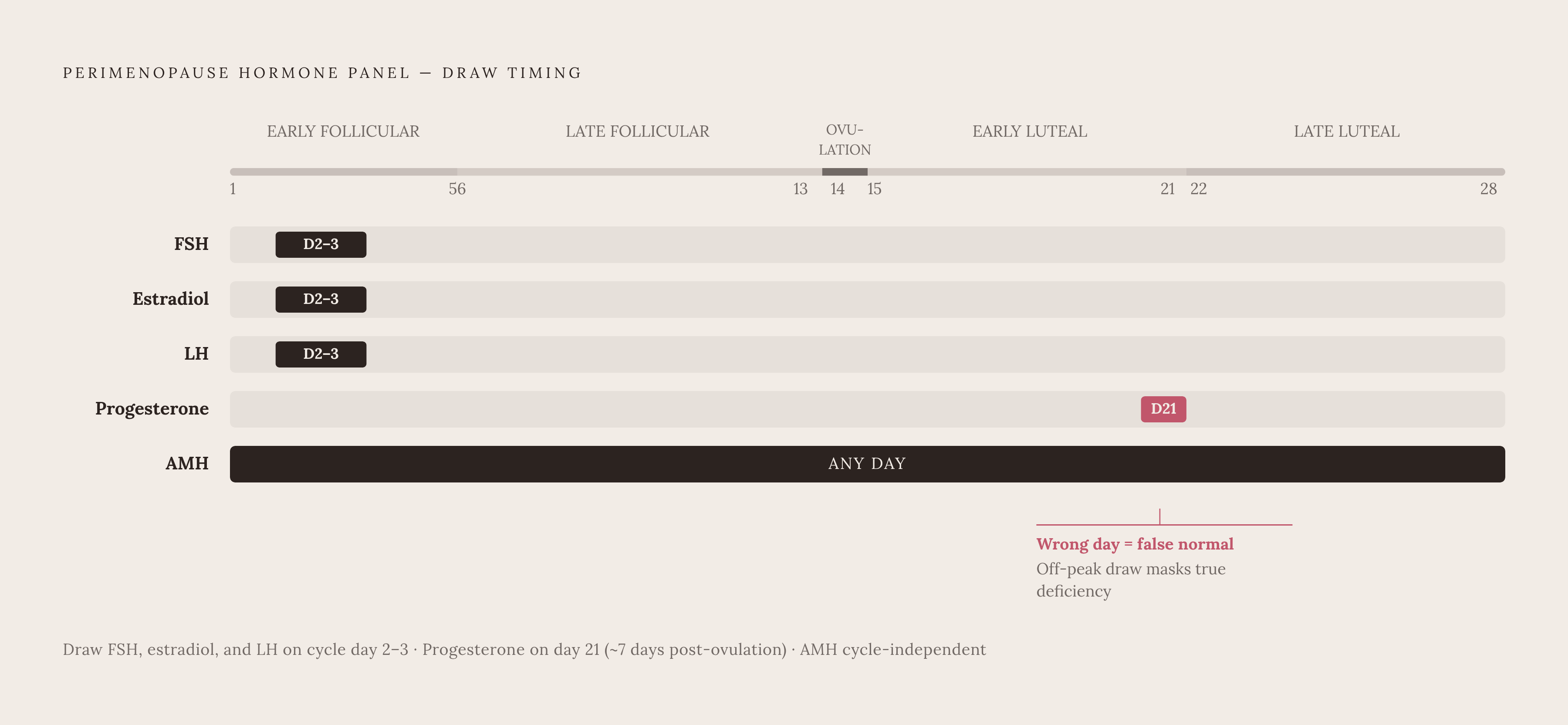
Timing decides whether a panel means anything at all, a fact central to the process rather than a minor scheduling detail bolted onto the end of it. Female hormones fluctuate throughout the cycle by design, rising and falling in a predictable rhythm while cycles are still regular. Test at the wrong point in that rhythm and a perfectly normal fluctuation can look like a red flag, or a genuine problem can hide behind a number that happens to look fine on that particular day. Get the day wrong and the whole panel becomes worse than useless, because a bad number now carries false confidence.
FSH, estradiol, and LH get drawn early in the follicular phase, typically day 2 or 3 of the cycle, while progesterone has to wait until the mid-luteal phase, roughly a week after ovulation, to mean anything at all. Once cycles start lengthening or skipping entirely, which is the norm rather than the exception in later perimenopause, that clean timing window gets harder to hit. AMH is the one core marker that sidesteps the problem entirely, since it can be tested on any day regardless of where a woman sits in her cycle. So what happens to the rest of the panel once cycles turn genuinely unpredictable? In practice it means retesting FSH weeks apart rather than trusting a single draw. A panel built on careful timing, read as a set rather than as isolated numbers, is what turns a pile of lab values into an actual picture of where a woman stands in the transition.
PCOS affects 10–13% of reproductive-aged women globally, according to the World Health Organization https://www.testing.com/tests/female-hormone-panel/. Between ages 40–44 and 55–59 years, median testosterone concentrations declined from 0.56 to 0.42 nmol/L https://www.ncbi.nlm.nih.gov/pmc/articles/PMC12552971/. Median androstenedione concentrations declined from age 40–44 to 65–69 years by 51% https://www.ncbi.nlm.nih.gov/pmc/articles/PMC12552971/. Median DHEA concentrations declined from age 40–44 to 65–69 years by 33% https://www.ncbi.nlm.nih.gov/pmc/articles/PMC12552971/. The reference range for FSH in a premenopausal woman in the early follicular phase is 3–10 mIU/mL https://holistic.health/journal/hormone-panel-explained-tests. The reference range for LH in a premenopausal woman in the early follicular phase is 2–10 mIU/mL https://holistic.health/journal/hormone-panel-explained-tests. The reference range for estradiol in a premenopausal woman in the early follicular phase is 25–75 pg/mL https://holistic.health/journal/hormone-panel-explained-tests. Oestradiol levels below 100 pmol/L in the early follicular phase suggest low ovarian output https://lolahealth.com/blogs/longevity/perimenopause-blood-test-guide. The normal TSH range is 0.27–4.2 mIU/L https://lolahealth.com/blogs/longevity/perimenopause-blood-test-guide. Many women feel best with TSH below 2.5 mIU/L https://lolahealth.com/blogs/longevity/perimenopause-blood-test-guide. The UK recommended minimum vitamin D level is 50 nmol/L https://lolahealth.com/blogs/longevity/perimenopause-blood-test-guide. Most specialists suggest 75–100 nmol/L is optimal for vitamin D https://lolahealth.com/blogs/longevity/perimenopause-blood-test-guide.