Perimenopause vs Insulin Resistance Diagnostic Criteria
Distinguishing these conditions requires menstrual history, hormone timing.
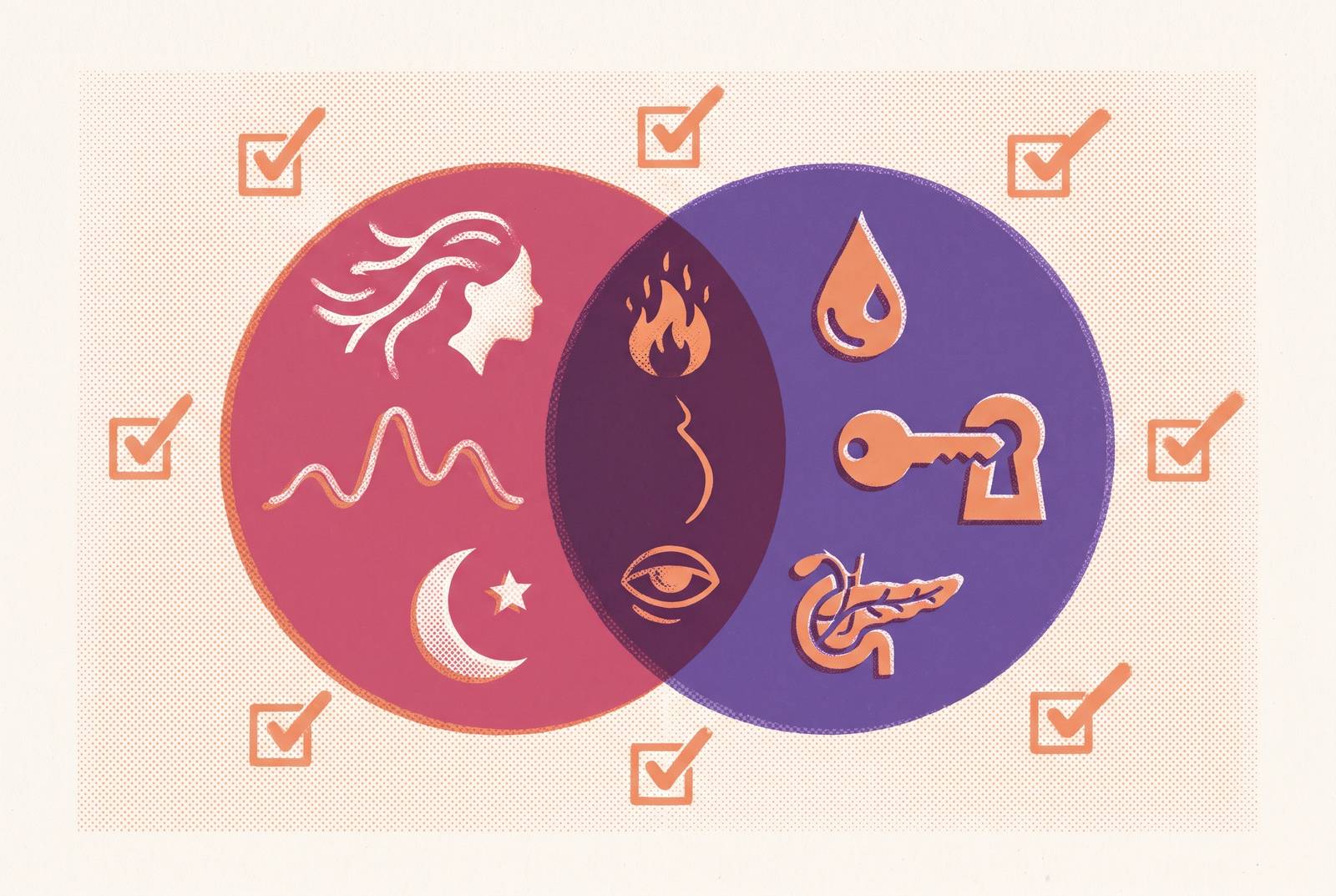
Perimenopause and insulin resistance can both cause weight gain, fatigue, brain fog, sleep disruption, and mood changes, making symptoms alone an unreliable way to distinguish them. Their diagnostic criteria differ, and both conditions can be present at once. Avoiding perimenopause misdiagnosis and dismissed symptoms requires interpreting menstrual history, appropriately timed hormone tests when indicated, and metabolic markers together rather than treating one normal result as the whole answer.
Why perimenopause and insulin resistance are so often confused for each other
A woman in her early-to-mid 40s presents with fatigue, unexplained weight gain, and mood shifts. Is the starting point reproductive aging, metabolic disruption, or a psychiatric assessment? Brain fog and disrupted sleep deepen the ambiguity because they belong to the shared symptom pattern.
Cycle irregularity, hot flashes, night sweats, vaginal dryness, and urogenital complaints point toward perimenopause, while pronounced carbohydrate cravings, post-meal energy crashes, and acanthosis nigricans suggest insulin resistance. Yet hot flashes and palpitations can resemble hypoglycemic episodes. Insulin resistance can also contribute to heavy periods through uterine lining growth and can suppress progesterone, complicating a distinction based on menstrual symptoms. When most of the appointment is spent on mood changes, anxiety or depression may be seen as the cause before hormonal change or insulin resistance is considered.
How declining estrogen during perimenopause directly drives insulin resistance
Estrogen improves insulin sensitivity, so its decline can reduce the body’s responsiveness to insulin. The overlap has a biological basis. Early perimenopause brings large estrogen fluctuations that can intermittently reduce insulin sensitivity, while later perimenopause brings a more sustained decline. Poor glycemic control is also associated with greater vasomotor symptom burden, so metabolic disruption may worsen hot flashes and night sweats attributed entirely to reproductive hormones.
As estrogen falls, loss of muscle mass, or sarcopenia, can become an independent risk factor for insulin resistance. When androgens are relatively high, they can further raise the risk of insulin resistance, which can in turn raise androgen levels. These reinforcing effects explain why identifying one condition does not exclude the other.
What the diagnostic criteria for perimenopause actually require — and what they miss
The Stages of Reproductive Aging Workshop framework, STRAW+10, uses menstrual changes to determine the stage of reproductive aging, with FSH levels helping to support that assessment. Early perimenopause involves persistent cycle irregularity of seven or more days from the usual cycle length. Late perimenopause involves a prolonged interval without menstruation. Postmenopause begins after 12 consecutive months without a period.
For women over 45 with typical symptoms, the prevailing clinical approach allows diagnosis without hormone testing. This recognizes that hormone levels fluctuate and that menstrual history can be more informative than an isolated result. When FSH is measured, a value above 30 U/L is consistent with perimenopause or menopause, but a lower value does not exclude perimenopause. A normal draw is not a veto. Pyne et al. 2024 also note that age alone has limited diagnostic usefulness because onset and symptom patterns vary.
Ovulation creates a further interpretive gap. A woman can have elevated estrogen yet remain symptomatic if she is not ovulating and therefore is not producing progesterone as expected. Progesterone testing needs a mid-luteal sample, approximately seven days after ovulation, with a level above 3 ng/mL confirming that ovulation occurred. AMH shows how much ovarian reserve remains, which is different from what hormones are doing that day. Younger age, a temporarily low FSH result, or apparently reassuring estrogen can each leave an incomplete picture.
What the diagnostic criteria for insulin resistance actually require — and where they fall short
There is no single diagnostic cutoff for insulin resistance that everyone agrees on. Thresholds vary with the population, assay, and clinical context, which limits how confidently one number can be interpreted. HOMA-IR, the Homeostasis Model Assessment of Insulin Resistance, is calculated as fasting insulin in µU/mL multiplied by fasting glucose in mmol/L, divided by 22.5. A 2023 retrospective analysis of women with PCOS identified a threshold of 2.1.
The more practical gap concerns what gets measured. Fasting glucose shows the blood sugar level, and fasting insulin helps show how much insulin the body needs to keep it there. Insulin can stay high while glucose is still within its reference range. Standard metabolic panels often omit fasting insulin entirely, leaving that compensatory effort unseen. HbA1c can also look normal even when insulin resistance is present.
A reassuring glucose result establishes the blood sugar level. Fasting triglycerides, HDL cholesterol, and waist circumference can add context because they contribute to the metabolic syndrome picture. However, metabolic syndrome and insulin resistance are not interchangeable diagnoses. The distinction prevents a broad risk profile from being mistaken for a direct measure of insulin sensitivity.
How the two diagnostic pictures overlap and where they diverge on a lab report
Rising or fluctuating FSH, declining AMH, variable estradiol, and mid-luteal progesterone below the ovulatory threshold can support concern about the reproductive transition. Each result needs to be read with its timing and context in mind. In particular, interpreting low progesterone as evidence of perimenopause requires additional findings, and estradiol variability is a pattern across draws. Cycle changes consistent with STRAW+10 connect the laboratory findings to reproductive staging.
The metabolic pattern asks a different question. Elevated fasting insulin, HOMA-IR above a context-appropriate threshold, elevated fasting triglycerides, low HDL, or rising HbA1c point toward metabolic disruption, including when cycles remain regular. Normal glucose cannot separate the conditions, while abdominal weight gain, poor sleep, and mood changes also lack that discriminatory value. Elevated testosterone or DHEA-S may bridge the two pictures rather than resolve them, because insulin resistance and androgen excess can reinforce one another.
Testing only hormones risks overlooking metabolic disruption, while testing only metabolic markers can leave hormonal change unexplored. Neither panel assigns every symptom to one cause. A single draw describes the sampled moment, whereas repeated, appropriately timed measurements can reveal the trajectory.
Conditions that further complicate the differential — thyroid dysfunction and PCOS
Thyroid dysfunction belongs in the differential because fatigue, weight gain, brain fog, mood changes, and irregular cycles can resemble either presentation. It needs consideration before those symptoms are attributed entirely to reproductive aging or insulin resistance. TSH is the standard thyroid screen. Free T4 helps make sense of the results, and some clinical protocols also use free T3 and thyroid antibodies. The purpose is not to expand testing indiscriminately, but to investigate another plausible explanation for the same complaints.
PCOS complicates both menstrual staging and metabolic interpretation. Irregular cycles and androgen excess are features of PCOS, and insulin resistance is present in a substantial proportion of affected women, although estimates vary. When a cycle is already irregular, it is harder to spot new changes linked to the stage of reproductive aging. Rotterdam, Androgen Excess Society, and NIH diagnostic criteria require exclusion of other endocrine disorders, including thyroid dysfunction, non-classical congenital adrenal hyperplasia, adrenal hyperandrogenism, and hyperprolactinemia. A woman with PCOS can also develop perimenopausal changes and insulin resistance simultaneously, so the history cannot be reduced to a choice between competing labels.
Why the standard clinical approach — no testing for women over 45 with typical symptoms — fails a significant portion of this population
The no-routine-hormone-testing approach answers a narrow question about recognizing typical perimenopause. It does not establish that every accompanying symptom has the same cause or remove the need to consider metabolic or thyroid disease. The distinction is easy to lose. When clinicians stop looking after making a clinical diagnosis, they may miss insulin resistance because its symptoms are so familiar. Conversely, the over-45 threshold can become an inappropriate barrier to considering reproductive changes in younger women.
Cunningham et al. 2025 surveyed 4,432 U.S. women and found that women aged 30–45 had symptoms that affected their lives and were seeking clinical help. That finding challenges age-based dismissal without proving that every symptom in that group is perimenopausal. Pyne and colleagues also describe women feeling that symptoms are not taken seriously and clinicians feeling inadequately trained to diagnose and treat perimenopause. A guideline limitation can therefore be compounded by a knowledge gap. Ordering only FSH, then interpreting a normal result as exclusion, offers apparent certainty without resolving the differential.
Mood symptoms stand out, but the hormonal and metabolic processes behind them are harder to see unless clinicians look for them. Treating those symptoms alone leaves the question of a physiological contributor unanswered. Understanding that gap gives a patient a concrete basis for asking what else has been considered.
What a complete diagnostic workup looks like when both conditions are on the table
In practice, a workup aimed at distinguishing these conditions should assess reproductive and metabolic evidence together, with the clinician selecting tests for the presentation. “Complete” should mean that the relevant diagnostic questions are addressed. Cycle history remains central to that assessment. FSH and estradiol can contribute when interpreted with their variability in mind, rather than treated as stand-alone confirmation or exclusion.
Timing is especially important for progesterone. A mid-luteal draw falls approximately on day 21 of a 28-day cycle, or seven days after confirmed ovulation. A low result needs that timing context before it can suggest anovulation. AMH adds information about ovarian reserve, particularly in younger women with ambiguous presentations, while testosterone and DHEA-S are relevant when androgenic symptoms are present. These markers answer distinct questions and should not be treated as substitutes for one another.
On the metabolic side, fasting insulin is the key addition when previous testing has measured only glucose or HbA1c. Fasting glucose permits the HOMA-IR calculation, and HbA1c adds glycemic context without excluding insulin resistance when normal. A HOMA-IR reference point above 2.0–2.1 is used in some settings, but requires population-appropriate interpretation. Fasting triglycerides and HDL help characterize the associated metabolic pattern. TSH, with free T4 where appropriate, addresses the thyroid alternative.
Follow-up changes what those results can mean. One normal FSH result differs from three draws across six months showing a rising trajectory. Likewise, a single HOMA-IR measurement cannot show how insulin sensitivity changes as estrogen falls. The value of repeat testing lies in answering an unresolved clinical question over time.
How to use this information when talking to a healthcare provider
For the clinical encounter, the most useful posture is to ask for an explanation that accounts for the whole pattern. Bring dated observations about symptoms, cycle length, missed periods, and flow changes. Include their context. Post-meal energy crashes and carbohydrate cravings raise a different question from night sweats or changing cycle intervals, even when they occur together. A record over months gives the clinician evidence that a single appointment cannot supply.
The discussion can then connect each proposed test to a purpose. Ask whether fasting insulin should be measured with fasting glucose, particularly if earlier reassurance rested on normal glucose or HbA1c. For progesterone, ask how the draw will be timed to ovulation. If a normal FSH result has been used to dismiss perimenopause, ask whether fluctuation warrants reassessment, and whether thyroid testing could clarify the picture.
If all symptoms are attributed to stress, anxiety, or age without considering hormonal or metabolic contributors, a second opinion from a clinician trained in perimenopause and metabolic health is reasonable. Ongoing symptom records and appropriately selected follow-up tests can show whether symptoms are worsening, stabilizing, or tracking a distinct metabolic process.